Spotlight On: Endometriosis

 This month we cast a spotlight on articles, SurgeryU videos, and Journal of Minimally Invasive Gynecology (JMIG) article recommendations from the AAGL Endometriosis Special Interest Group (SIG) led by Chair, Maria Victoria Vargas, MD, MS.
Access to SurgeryU and JMIG are two of the many benefits included in AAGL membership. The SurgeryU library features high-definition surgical videos by experts from around the world. JMIG presents cutting-edge, peer-reviewed research, clinical opinions, and case report articles by the brightest minds in gynecologic surgery.
SurgeryU video recommendations by our SIGs are available for public access for a limited time. The links to JMIG article recommendations are accessible by AAGL members only. For full access to SurgeryU, JMIG, CME programming, and member-only discounts on meetings, join AAGL today!

SIG Recommended SurgeryU Video #1:
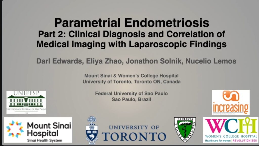
Parametrial Endometriosis Part 2: Clinical Diagnosis and Correlation of Medical Imaging with Laparoscopic Findings
By Darl Edwards MD, Eliya Zhao MD, Meir J. Solnik, MD, and Nucelio Lemos, MD
This is the second video from a practical series about parametrial endometriosis developed by surgical teams from Toronto and São Paulo. This second video describes how parametrial endometriosis can present clinically with neuropathic pain not typically associated with gynecologic disease. The video further correlates imaging findings of parametrial endometriosis to the surgical appearance of the disease.
 Click Image to View Video
SIG Recommended SurgeryU Video #2:
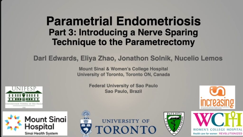
Parametrial Endometriosis Part 3: Introducing a Nerve Sparing Technique to the Parametrectomy
This is the third video from a series of videos about endometriosis of the parametria. This video reviews the parametrial anatomy and displays a structured ultra lateral nerve sparing technique to excise parametrial endometriosis. The video then goes on to summarize the data collected on outcomes of this technique at a high-volume center.

JMIG Article Recommendation #1:
Impact of Surgery for Deep Infiltrative Endometriosis Before In Vitro Fertilization: A Systematic Review and Meta-Analysis
Gemma Casals, MD, PhD, María Carrera, MD, José Antonio Domínguez, MD, PhD, Mauricio Simões Abrão, MD, PhD, Francisco Carmona, MD, PhD
This article by Dr. Casals and colleagues is a systematic review of the impact of surgery for deep endometriosis prior to IVF. The collated data showed a benefit in live birth rates for surgical excision of deep endometriosis of the digestive tract prior to IVF (OR2.43; 95% CI 1.13-5.22). The results are compelling but there were no randomized controlled trials included and there were minimal results regarding complications resulting from surgery. However, this systematic review does support the notion that strategic complete surgical excision of endometriosis of the digestive tract may be of benefit for patients with infertility in conjunction with ART.
JMIG Article Recommendation #2:
Pregnancy Outcomes After Uterus-Sparing Operative Treatment for Adenomyosis: A Systematic Review and Meta-Analysis
Lijuan Jiang, MM, Yue Han, MM, Zixuan Song, MD, and Yan Li, MD, PhD
This article is a meta-analysis which reviews pregnancy outcomes for patients with infertility who undergo fertility sparing surgical excisional or nonexcisional (UAE, HIFU, MWA and RFA) treatment of adenomyosis. Although the current literature is not conclusive, this article suggests instances where these techniques might be beneficial for patients suffering from adenomyosis who desire fertility.

Most Difficult Case Video Article: New to SurgeryU!
A Challenging Case Due to Previous Incomplete Excision of Endometriosis
This video highlights the impact that incomplete surgery can have on patients with advanced endometriosis. This patient had a prior left ovarian cystectomy with extensive lysis of adhesions and left ureterolysis, but no excision of her extraovarian endometriosis. This is a video of a subsequent surgery to remove the remainder of her pelvic endometriosis, showing the extensive fibrosis and adhesive disease that was likely worsened by her prior surgery, creating an added challenge to removing her disease while preserving her fertility.
 Click Image to View Video
About the Author:
Maria Victoria Vargas, MD, MS, FACOG
Dr. Vargas is Chair of the AAGL Endometriosis Special Interest Group and Director of Minimally Invasive Gynecologic Surgery at Sibley Memorial Hospital, Johns Hopkins Medicine, in Washington, D.C.

Bridging Surgery with Cutting-Edge Single-Cell and Spatial Omics: Understanding Endometriosis Through Translational Research
Endometriosis is a complex, heterogenous disease for which we have little understanding of its basic pathophysiology. Different phenotypes of endometriosis cause a range of symptoms from “no” symptoms to debilitating pain and/or infertility. Basic scientists are utilizing cutting edge technology to try to understand the mechanisms of endometriosis using tissue obtained from patients having surgery by advanced endometriosis surgeons. This article is going to briefly explain one of these technologies and then summarize some of the recent literature on endometriosis research.
Initial translational research in endometriosis used bulk RNA seq which was able to see what cells composed the lesions but not the ratio. Single cell technology allows the scientist to look at the ratio of different cells in the tissues. Spatial transcriptomics allows scientists to not only see what cells are in the tissue but also where they are within the lesion and how the cells interact with each other. Image 1 depicts these technologies using an analogy with Legos and figures from a study by Tan et al in Nature Cell.[1]
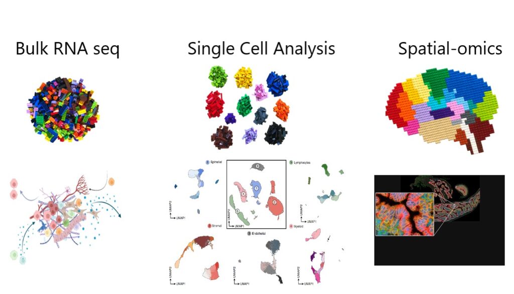 Image 1
The study by Tan et al demonstrated distinct differences in ratios of cells seen in patients with and without endometriosis. Epithelial cells made up the bulk of the tissue in control endometrium where fibroblasts and immune cells (specifically T lymphocytes and NK cells) made up the bulk of the cells seen in endometriosis eutopic endometrium. Epithelial cells were also minor components of the endometriotic lesions, where stromal fibroblasts, endothelial and immune cells made up >70% of the lesion. When looking at the spatial-omics, the immune cells as well as fibroblasts in endometriosis endometrium and lesions expressed different genes than cells in control endometrium.1
Fonseca et al published an atlas of cell types in endometriosis lesions and saw differences not only in cell populations but in transcriptomic signatures between peritoneal lesions and endometrioma supporting the phenotypic differences seen not only on gross inspection but in behavior between peritoneal and ovarian lesions.[2]
A review by Nezhat recently published in NPJ Women’s Health summarized the recent literature on endometriosis translational research including single cell analysis, proteomic and metabolomic studies, and genomic studies. They emphasized the importance of surgeons and basic scientists to work together to better understand endometriosis.[3] The aspiration is to find a non-invasive diagnostic test and targeted therapies for endometriosis which will come from the collaboration of basic science technology, medicine, and surgery.
References:
- Tan Y, Flynn WF, Sivajothi S, Luo D, Bozal SB, Dave M, Luciano AA, Robson P, Luciano DE, Courtois ET. Single-cell analysis of endometriosis reveals a coordinated transcriptional programme driving immunotolerance and angiogenesis across eutopic and ectopic tissues. Nature Cell Biology 2022; 24(8):1306-18.
- Fonseca MAS, et al. Single-cell transcriptomic analysis of endometriosis Nat Genet 2023; 55:255-67.
- Nezhat CR, Oskotsky TT, Robinson JF, Fisher SJ, Tsuei A, Liu B, Irwin JC, Gaudilliere B, Sirota M, Stevenson DK, Giudice LC. Real World Perspectives on Endometriosis disease phenotyping through surgery, omics, heath data, and artificial intelligence. NPJ women’s health 2025 3(8): 1-16.
About the Author:
Danielle Luciano, MD

Dr. Luciano is a Board Member of the AAGL Endometriosis and Reproductive Surgery SIG, Professor and Interim Chair of the Obstetrics and Gynecology and Co-Director of CTEndoRISE at UConn Health, University of Connecticut in Farmington, Connecticut.