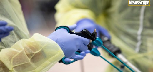
Popular Difficult Case Series Continues

Articles featured in this month’s Difficult Series were submitted by the Fibroids SIG and Pediatric Adolescent Gynecology SIG. Thank you to all the AAGL SIG chairs, vice-chairs, and members – for the informative articles and for supporting this popular series and special thanks to the Fibroids and PAG SIG for these submissions!
Fibroids SIG Difficult Case: Large Uterine Fibroid Causing Bulk Symptoms
A 37-year-old G2P2002 presented for a second opinion regarding management of a large uterine fibroid causing bulk symptoms. In her first pregnancy she had a “dime-sized” fibroid which had grown to a 15-centimeters at the time of her repeat cesarean delivery.
Following delivery, a palpable mass above the level of the umbilicus was persistent. A pelvic ultrasound and MRI showed a normal-sized uterus with a 25 cm x 18 cm x 11 cm exophytic heterogenous fibroid. It extended from the left posterior uterus on a thick stalk and displaced the uterus and bladder anteriorly and to the right. There was no evidence of hydronephrosis. The patient remained mildly symptomatic from the fibroid, complaining only of pain when standing for long periods of time. She was recommended to have an abdominal hysterectomy via midline vertical incision. However, she hoped to maintain her fertility.
On our initial exam the mobile fibroid was palpated 3 cm above the umbilicus and extended the width of her abdomen. An endometrial biopsy showed proliferative endometrium. A 3-month depot of GnRH agonist was given to shrink the fibroid. She was scheduled for a laparoscopic, possible abdominal, myomectomy. Pre-operative exam revealed that the fibroid was now 2 cm below the umbilicus with the same width.
Rectal misoprostol, dilute vasopressin and IV tranexamic acid were used intraoperatively to minimize bleeding. A Palmer’s Point entry was used to avoid uterine trauma. The uterus was adhered to the anterior abdominal wall. The fibroid arose from the left posterior uterus and was completely contained within the broad ligament.
We used a blunt probe to push the fibroid medially and identified the left ureter in the retroperitoneal space lateral to the IP ligament. A horizontal incision was made over the fibroid in the posterior broad ligament, and we enucleated the fibroid with blunt dissection and ultrasonic energy. Once free from its retroperitoneal attachments, the fibroid was mobilized into the upper abdomen and its pedicle to the posterior uterus was well-visualized. We desiccated and transected the pedicle with ultrasonic energy. The posterior peritoneal defect was oversewn with barbed suture. The fibroid was placed in a large tissue extraction bag and morcellated via an umbilical mini-laparotomy incision. Estimated blood loss for the procedure was 200 cc. The patient was monitored for dizziness in the PACU and discharged from the hospital on post-op day #1.
Large broad ligament fibroids can be removed laparoscopically with uterine preservation. Consider GnRH agonists to decrease fibroid bulk pre-operatively. If possible, early identification of the ureters is key to avoiding injury. There is no myometrium in the broad ligament and no pseudo-capsule. As broad ligament fibroids grow, they push the ureter and vessels away. The peritoneum overlying the fibroid is translucent and you can make an incision where the ureter is not visible. While putting traction on the fibroid, the adventitia is pushed off the fibroid, thus sweeping the ureter and vessels out of harms-way.
We all encounter fibroid problems. This NewsScope issue, features articles by Fibroids Special Interest Group (FSIG) members to help expand your knowledge of the treatment of fibroids. In addition, two SurgeryU videos accompany their didactic. Dr Malhotra helps us visualize laparoscopic myomectomy for a broad ligament fibroid. Dr Musselman et al demonstrate how pre-op assessment is important and led to combined hysteroscopic resection and laparoscopic radio frequency ablation.
Kristen Pepin, MD, MPH, Assistant Professor of Obstetrics and Gynecology, Weill Cornell Medicine, New York Presbyterian Hospital

PAG SIG Difficult Case: Vaginal Bleeding in a Prepubertal Patient
A 7-year-old healthy female presented with new onset vaginal bleeding. Review of systems was otherwise unremarkable. Physical exam and initial labs confirmed no evidence of pubertal development. A pelvic ultrasound revealed small ovaries bilaterally and a prepubertal uterus measuring 4.1 x 2.1 x 2.7 cm with an endometrial polypoid mass, 1.6 x 1.2 x 1.7 cm with vascularity at its base. Magnetic resonance imaging (MRI) revealed a 1.6 cm bilobed hypervascular enhancing polypoid lesion in the endometrium with fluid distention of the endometrial cavity concerning for sarcoma versus atypical endometrial polyp.
The patient was taken for vaginoscopy, hysteroscopy and endometrial biopsy. We anticipated challenges with her hypoestrogenic genital tract, limited working space and optimal equipment to biopsy and/or resection of the endometrial mass. A rigid, continuous flow, 9.5 Fr pediatric offset operative cystoscope with a 6 Fr working channel using 1.5% glycine solution as a distension media under positive pressure was chosen. The scope was advanced through the hymen and the labia majora pinched to allow fluid distention of the vagina. A global view of vagina and cervix revealed typical prepubertal appearance without lesions. The cystoscope was unable to be advanced beyond her internal cervical os. The scope was removed, and the cervix was visualized by applying retraction with the disarticulated upper and lower blades of a small Pederson speculum. The anterior lip of the cervix wad grasped with an allis tenaculum and the cervix gently dilated to a 4mm Hegar dilator to allow the cystoscope to advance. Intrauterine placement of the cystoscope was confirmed with visualization of tubal ostia and the bilobed polypoid mass was noted. The cystoscope was removed and the 9 Fr cysto-resectoscope was introduced. The resectoscope provided a limited visual field, so was removed. The TruClear ™ 5.0 mm system was considered for resection. Given the larger diameter needed for entry and long working channel length, it was not optimal for her small cervical os and limited vaginal depth, as such it was not used. As the priority in this case was for tissue diagnosis, the 9.5 Fr cystoscope was reintroduced. The biopsy forceps was inserted through the working channel and samples of the lesions were taken under direct visualization. The bugbee electrode was inserted through the working channel and used for hemostasis where needed. Once hemostasis was confirmed all instruments were removed with ease. Her pathology returned congenital hemangioma.
This case posed a particular challenge given the limitations for operative hysteroscopic access in a prepubertal patient. The need for assessment of the endometrial cavity is rare in this age group, therefore there is limited and variable clinical practice and published reports limited to postmenarhcal patients. (1) Although access to the endometrial cavity may be obtained with smaller diameter endoscopes, there are significant limitations for adequate sampling or resection of endometrial lesions.
Gylynthia E. Trotman, MD, MPH, FACOG is Chief of Pediatric and Adolescent Gynecology, Assistant Professor of Obstetrics and Gynecology and Assistant Professor of Pediatrics at Icahn School of Medicine at Mount Sinai Health System/Kravis Children’s Hospital

- Jolinda Johary et.al. Use of hysteroscope for vaginoscopy or hysteroscopy in adolescents for the diagnosis and therapeutic management of gynecologic disorders: a systematic review. J Pediatr Adolesc Gynecol. 2015 Feb;28(1):29-37


